Graphiola Leaf Spot
|
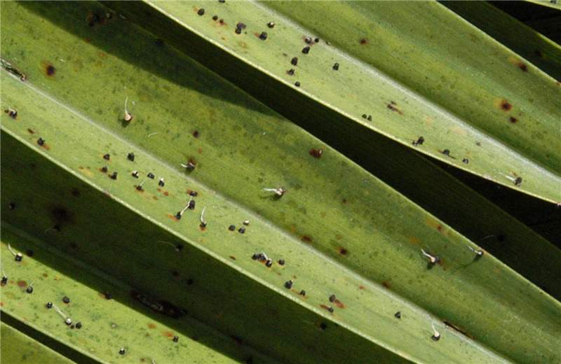
Figure 2. Palm leaflets with numerous sori and emerging spores. Note that this leaf does not have potassium deficiency. There are no leaf spot symptoms, only the sori of the fungus. Photo by M. L. Elliott.
|
|
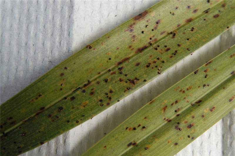
Figure 1. Sori of Graphiola phoenicis have erupted through the leaf epidermis. Some of the sori have filaments emerging from them. The mostly brown spots are not a symptom of Graphiola leaf spot, but a symptom of potassium deficiency. Photo by M. L. Elliott.
|
|
Figure 4. The brown spots are not early symptoms of Graphiola leaf spot, but are symptoms of potassium deficiency. Photo by M. L. Elliott.
|
|
Figure 3. Palm leaflets with numerous sori and emerging filaments (look like tiny strings) with spores. There are no leaf spot symptoms, only the sori of the fungus. Photo by M. L. Elliott.
|
|
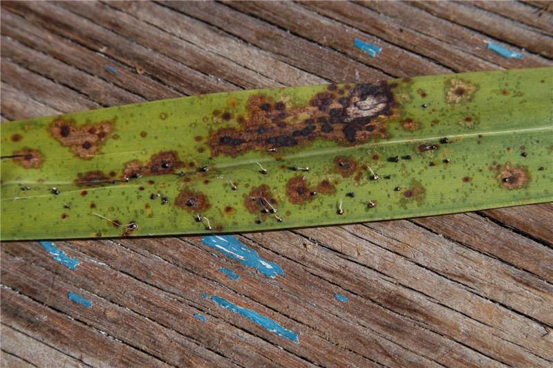
Figure 5. A leaf affected by both Graphiola leaf spot and Stigmina leaf spot. Signs of Graphiola phoenicis are the small back bodies (sori), many with filaments emerging from the sori. Stigmina palmivora symptoms are the large brown spots with dark edges and darker but flat centers. In some cases, a G. phoenicis sorus is superimposed upon the S. palmivora leaf spot. Photo by M. L. Elliott.
|
Other common names
false smut
Scientific name of pathogen
Graphiola phoenicis: Kingdom Fungi, Phylum Basidiomycota
Hosts
Graphiola phoenicis has been reported on numerous palm species, but is most commonly associated with Phoenix canariensis and Phoenix dactylifera.
Other palms on which G. phoenicis has been observed include: Acoelorrhaphe wrightii, Arenga pinnata, Butia capitata, Chamaerops humilis, Coccothrinax argentata, Cocos nucifera, Dypsis lutescens, Livistona alfredii, Livistona chinensis, Phoenix loureirii, Phoenix paludosa, Phoenix roebelenii, Phoenix sylvestris, Phoenix theophrasti, Prestoea acuminata, Roystonea regia, Sabal minor, Sabal palmetto, Syagrus romanzoffiana, Thrinax morrisii, and Washingtonia robusta.
Distribution
The pathogen has a worldwide distribution.
Symptoms/signs
With Graphiola leaf spot, the signs of the disease are more prevalent and more easily observed than the symptoms of the disease. Both signs and symptoms will be observed on the oldest leaves, which are the lowest leaves in the canopycanopy:
the cluster of leaves borne at the tip of the stem
.
The initial symptoms of the disease are very tiny (1/32 inch or less) yellow or brown or black spots on both sides of the leaf bladeleaf blade:
the broad, flattened distal portion of a leaf
. They are easily missed without close observation. The fungus will emerge from these spots, rupturing the leaf epidermis (leaf surface). It is the resulting fungal reproductive structures (sori) that are most commonly observed, and which obscure any true symptoms (Fig. 1).
The sorus (sori is the plural form) is a fruiting body that is less than 1/16 inch in diameter. As the sorus matures and yellow spores are produced, short, light-colored filaments (thread-like structures) will emerge from the body, and the sorus becomes black (Figs. 2 and 3). These filaments aide in spore dispersal. Once the spores are dispersed, the sori deflate and appear like a black, cup-shaped body or black crater. You can easily see the sori, but you can also feel the sori with your finger as they are raised above the leaf epidermis. The number of sori indicates the level of infection.
This is a disease that can be easily identified by examining the leaf, as the fungus is easily observed with the unaided eye. A simple magnifying glass will provide adequate "close-up" views.
May be confused with
Graphiola leaf spot is often confused with potassium deficiency. However, the initial leaf spot symptoms of Graphiola leaf spot are minute in comparison to those associated with potassium deficiency. Furthermore, if a palm is exhibiting potassium deficiency, Graphiola leaf spot can be superimposed on the potassium deficiency (Figs. 1 and 4).
It is quite possible to observe Graphiola leaf spot and another leaf spot disease occurring on the same leaf and to observe the sori of Graphiola phoenicis occurring within the symptomatic leaf spot of the other disease (Fig. 5).


