Gallery
Texas Phoenix Palm Decline
Figure 6. The spear leaf has died and is hanging down below the canopy of this Phoenix sylvestris. Photo by M. L. Elliott.
Figure 6. The spear leaf has died and is hanging down below the canopy of this Phoenix sylvestris. Photo by M. L. Elliott.
Texas Phoenix Palm Decline
Figure 5. The second Phoenix dactylifera from the right has more dead leaves than the remaining healthy palms. Photo by B. Dick, City of Lakeland, Florida.
Figure 5. The second Phoenix dactylifera from the right has more dead leaves than the remaining healthy palms. Photo by B. Dick, City of Lakeland, Florida.
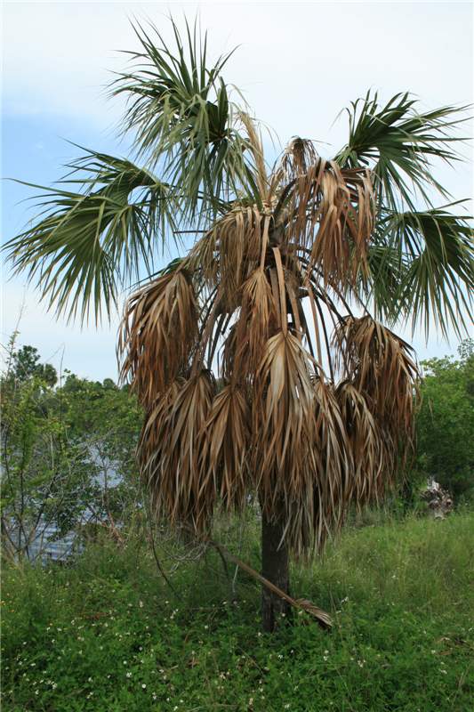 Figure 8. Sabal palmetto with two-thirds of the lower leaves necrotic and a dead spear leaf. Note that the next youngest leaves are still green. Photo by N. A. Harrison, University of Florida.
Texas Phoenix Palm Decline
Figure 8. Sabal palmetto with two-thirds of the lower leaves necrotic and a dead spear leaf. Note that the next youngest leaves are still green. Photo by N. A. Harrison, University of Florida.
Figure 8. Sabal palmetto with two-thirds of the lower leaves necrotic and a dead spear leaf. Note that the next youngest leaves are still green. Photo by N. A. Harrison, University of Florida.
Texas Phoenix Palm Decline
Figure 7. This photograph illustrates both a dying spear leaf and fruit stalks that have lost all of their fruit. Photo by M. L. Elliott.
Figure 7. This photograph illustrates both a dying spear leaf and fruit stalks that have lost all of their fruit. Photo by M. L. Elliott.
Thielaviopsis Trunk Rot
Figure 2. Syagrus romanzoffiana trunk collapse due to Thielaviopsis trunk rot. Only a few young green leaves remain. Note the "stem bleeding" or oozing of plant liquid down the trunk from the point of collapse.
Figure 2. Syagrus romanzoffiana trunk collapse due to Thielaviopsis trunk rot. Only a few young green leaves remain. Note the "stem bleeding" or oozing of plant liquid down the trunk from the point of collapse.
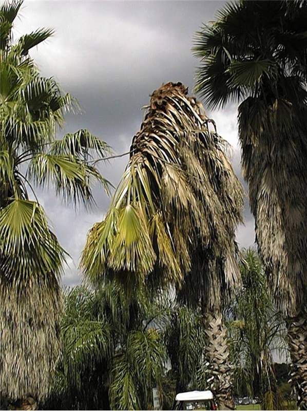 Figure 1. Trunk collapse of a Washingtonia robusta due to Thielaviopsis trunk rot. Note that most of the canopy was still green. Photo by F. W. Howard, University of Florida.
Thielaviopsis Trunk Rot
Figure 1. Trunk collapse of a Washingtonia robusta due to Thielaviopsis trunk rot. Note that most of the canopy was still green. Photo by F. W. Howard, University of Florida.
Figure 1. Trunk collapse of a Washingtonia robusta due to Thielaviopsis trunk rot. Note that most of the canopy was still green. Photo by F. W. Howard, University of Florida.
Thielaviopsis Trunk Rot
Figure 4. Only one side of this trunk has significant rot due to Thielaviopsis paradoxa. The fungus rots the trunk tissue from the outside to the inside. Photo by M. L. Elliott.
Figure 4. Only one side of this trunk has significant rot due to Thielaviopsis paradoxa. The fungus rots the trunk tissue from the outside to the inside. Photo by M. L. Elliott.
Thielaviopsis Trunk Rot
Figure 3. The three Cocos nucifera on the left have died from Thielaviopsis trunk rot. The palm in the foreground exhibits trunk collapse. Photo by M. L. Elliott.
Figure 3. The three Cocos nucifera on the left have died from Thielaviopsis trunk rot. The palm in the foreground exhibits trunk collapse. Photo by M. L. Elliott.
Thielaviopsis Trunk Rot
Figure 6. An example of "stem bleeding" on a Cocos nucifera trunk. The top of the blackened area was very soft and could be easily pushed in with the fingers. Photo by M. L. Elliott.
Figure 6. An example of "stem bleeding" on a Cocos nucifera trunk. The top of the blackened area was very soft and could be easily pushed in with the fingers. Photo by M. L. Elliott.
Thielaviopsis Trunk Rot
Figure 5. The canopy of Cocos nucifera in the center is wilted and necrotic due to a trunk infection by Thielaviopsis paradoxa. The infection site was just below the oldest leaf base. Photo by M. L. Elliott.
Figure 5. The canopy of Cocos nucifera in the center is wilted and necrotic due to a trunk infection by Thielaviopsis paradoxa. The infection site was just below the oldest leaf base. Photo by M. L. Elliott.
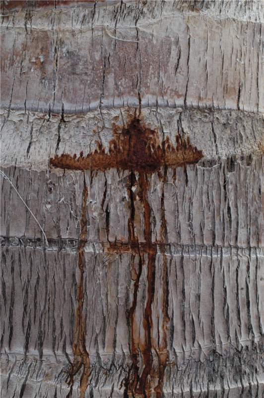 Figure 7. The trunk of this Cocos nucifera was just beginning to exhibit quot;stem bleedingquot;, but the large rusty-brown area at the top was already soft. Photo by M. L. Elliott.
Thielaviopsis Trunk Rot
Figure 7. The trunk of this Cocos nucifera was just beginning to exhibit "stem bleeding", but the large rusty-brown area at the top was already soft. Photo by M. L. Elliott.
Figure 7. The trunk of this Cocos nucifera was just beginning to exhibit "stem bleeding", but the large rusty-brown area at the top was already soft. Photo by M. L. Elliott.
Thrips Damage
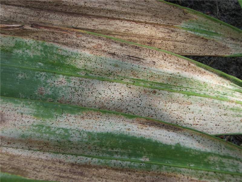
Figure 1. Lower surface of Veitchia sp leaf damaged by thrips feeding. Photo by T.K. Broschat
Figure 1. Lower surface of Veitchia sp leaf damaged by thrips feeding. Photo by T.K. Broschat
Thrips Damage
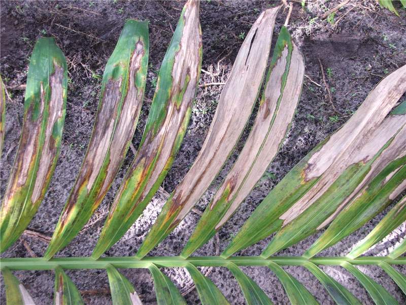
Figure 2. Upper surface of leaf of Veitchia sp. damaged by thrips feeding. Photo by T.K. Broschat
Figure 2. Upper surface of leaf of Veitchia sp. damaged by thrips feeding. Photo by T.K. Broschat
Water Stress
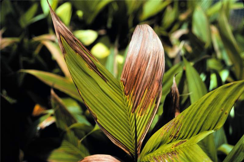
Figure 1. Leaf tip necrosis caused by water stress in Geonoma sp. Photo by T.K. Broschat.
Figure 1. Leaf tip necrosis caused by water stress in Geonoma sp. Photo by T.K. Broschat.
Water Stress
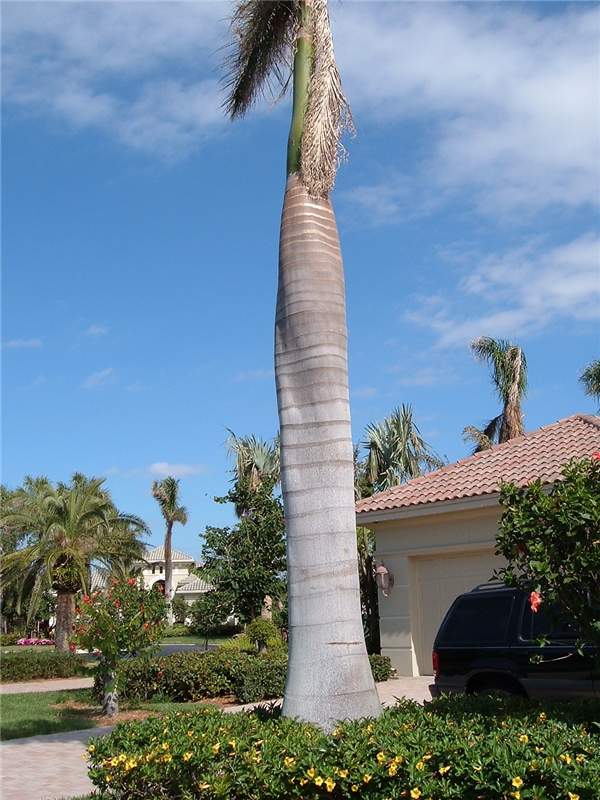
Figure 2. Collapsed and twisted trunk of water-stressed Roystonea regia. Photo by T.K. Broschat.
Figure 2. Collapsed and twisted trunk of water-stressed Roystonea regia. Photo by T.K. Broschat.
Wind Damage
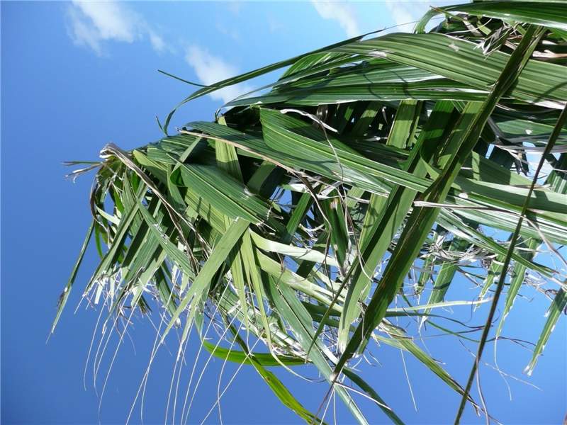
Figure 1. Tattered leaflets in Roystonea regia caused by high winds. Photo by T.K. Broschat
Figure 1. Tattered leaflets in Roystonea regia caused by high winds. Photo by T.K. Broschat
Wind Damage
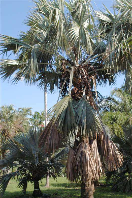
Figure 2. Broken leaves in Bismarckia nobilis caused by high winds. Photo by T.K. Broschat
Figure 2. Broken leaves in Bismarckia nobilis caused by high winds. Photo by T.K. Broschat


