Brevipalpus cuneatus
|
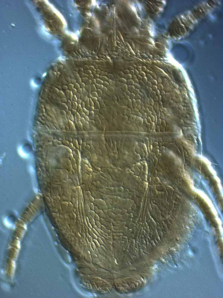
Fig. 1. Brevipalpus cuneatus female dorsum. |
|
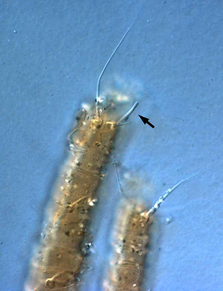
Fig. 2. Brevipalpus cuneatus female legs I-II - arrow indicates long solenidion. |
|
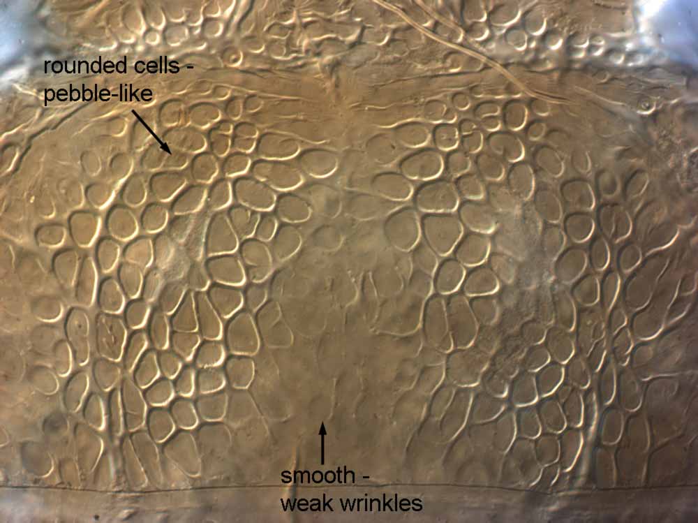
Fig. 2. Brevipalpus cuneatus female prodorsum. |
|
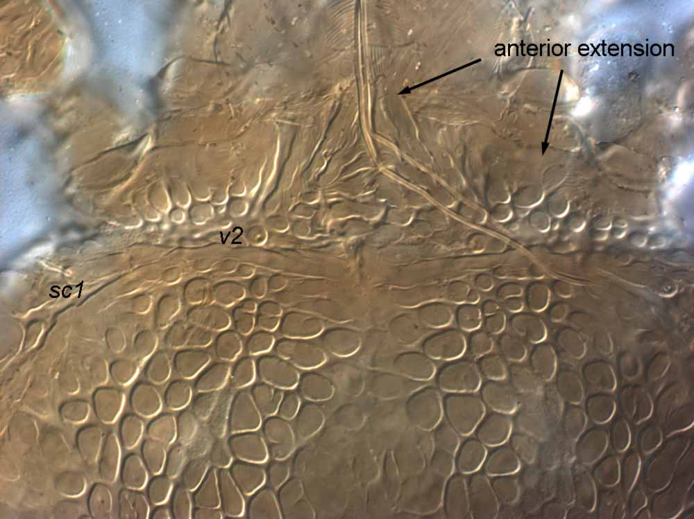
Fig. 4. Brevipalpus cuneatus female anterior extension of prodorsum. |
|
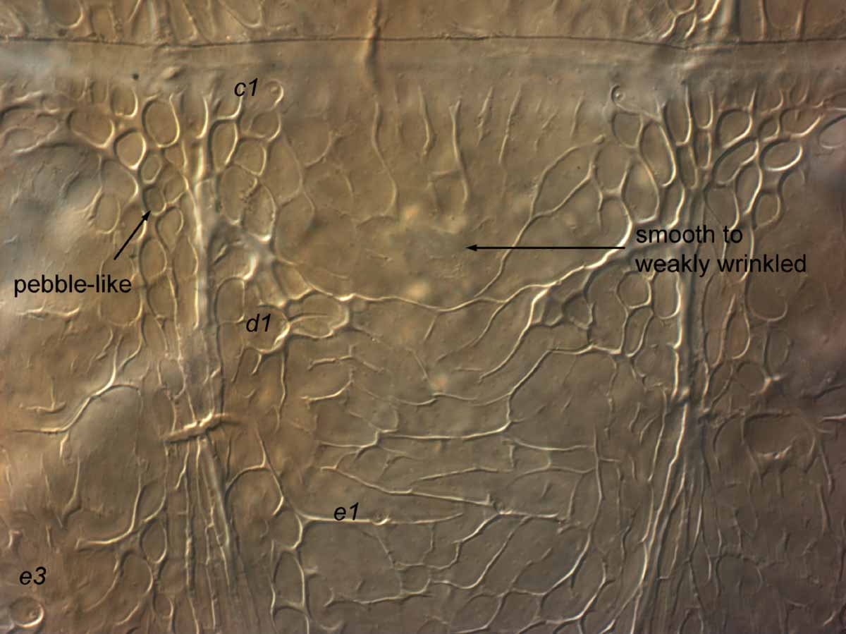
Fig. 5. Brevipalpus cuneatus female dorsal opisthosoma. |
|
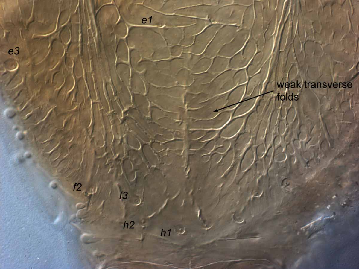
Fig. 6. Brevipalpus cuneatus female posterior dorsum. |
|
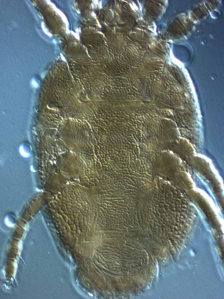
Fig. 7. Brevipalpus cuneatus female venter. |
|
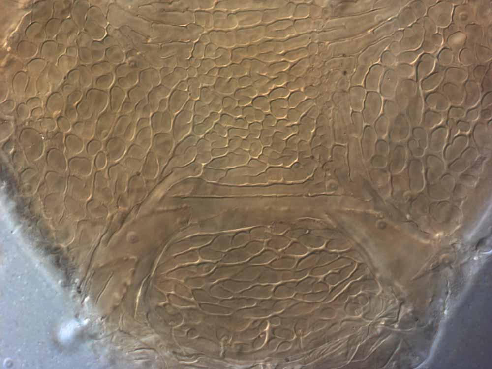
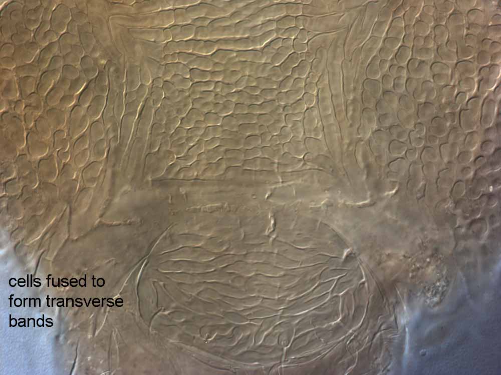
Fig. 8. Brevipalpus cuneatus female posterior venter - note lack of well defined ventral plate. |
|
Fig. 9. Brevipalpus cuneatus female posterior venter - note lack of well defined ventral plate. |
|
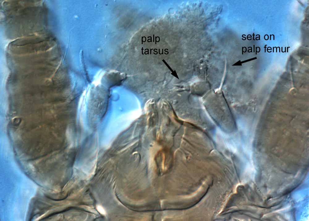
Fig. 10. Brevipalpus cuneatus female palp, indicating small palp tarsus, and seta on palp femur. |
|
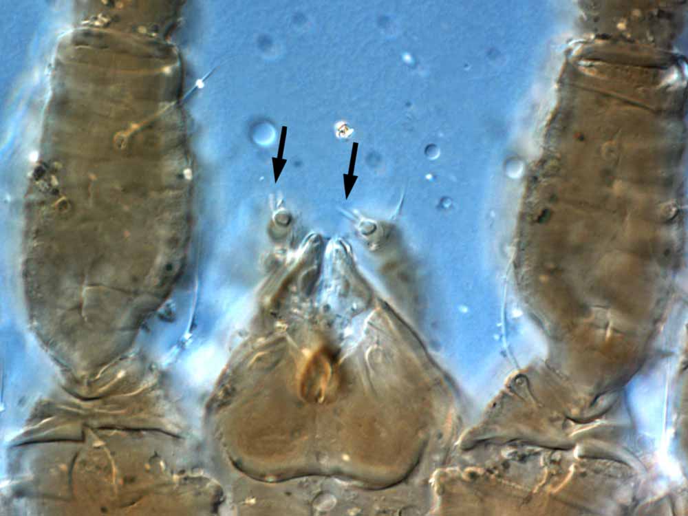
Fig. 11. Brevipalpus cuneatus female palp - arrow indicates 2 setae on palp tarsus. |
|
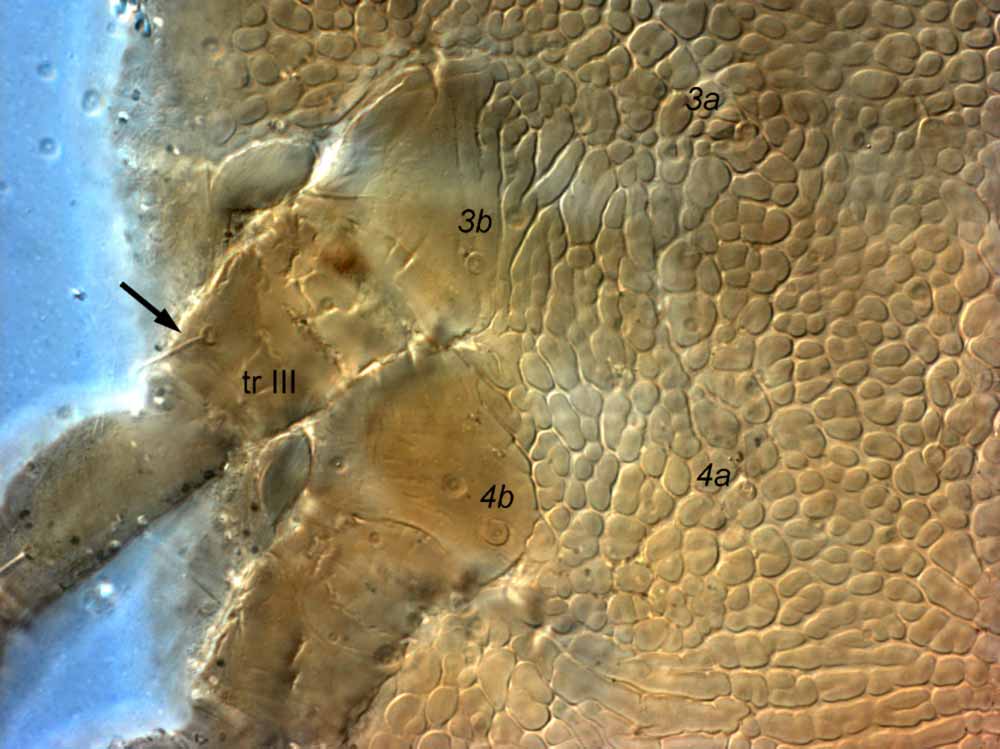
Fig. 12. Brevipalpus cuneatus female venter, arrow indicates seta on trochanter III (= tr III); note also small setae 3a, 4a. |
Authority
(Canestrini and Fanzago)
Taxonomic history
Caligonus cuneatus - original designation
Tenuipalpus cuneatus - Berlese (1887)
Brevipalpus cuneatus - Baker (1949)
Hystripalpus cuneatus - Mitrofanov & Strunkova (1979)
Species group characters
B. cuneatus species group (sensu Baker & Tuttle 1987) = f2 present; tarsus II with 1 solenidion; dorsal central setae (c1, d1, e1) different shape to dorsal lateral setae (c3, d3, e3); palp 4-segmented with 2 distal setae
Characters
- opisthosomal setae f2 present (= 7 setae around opisthosomal margin) (Figs. 1, 6)
- tarsus II with 1 solenidion distally (antiaxial); solenidia on tarsus I-II long (Fig. 2)
- prodorsum smooth centrally, with some weak wrinkles; with rounded closed cells laterally, forming strong pebble-like reticulation (Figs. 3, 4)
- anterior projection of prodorsum with strong pebble-like reticulation (Fig. 4)
- dorsal opisthosoma cuticle between c1-c1 and e1-e1 smooth to weakly wrinkled (rugose) (Fig. 5); cuticle laterad c1 with pebble-like reticulation; cuticle posterior e1-e1 with weak transverse folds and large rounded cells (Figs. 5, 6)
- seta f3 inserted off the body margin, medad f2 (Fig. 6)
- almost the entire ventral surface of body with strong pebble-like reticulation (Fig. 7)
- ventral plate not well defined, with small to medium circular cells (Figs. 8, 9)
- genital plate with large rounded cells transversely aligned (Figs. 8, 9), cells often fused to form transverse bands/folds (Fig. 9)
- spermatheca not visible
- palp tibia with protuberance; seta on femur strong, tapered, lightly barbed (Fig. 10)
- palp tarsus very small, with 2 setae - 1 solenidion, 1 eupathidion (Figs. 10-11)
- trochanter III with 1 seta (Fig. 12)
- ventral setae 3a and 4a very short (Fig. 12)
Distribution based on confirmed specimens
Italy (holotype)
Hosts based on confirmed specimens
unidentified hedge plant (holotype)
References
Baker (1949); Berlese (1887); *Canestrini & Fanzago (1876); Mitrofanov & Strunkova (1979)
* - original description

