

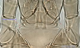
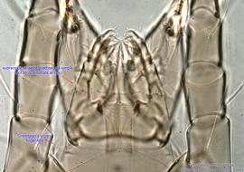
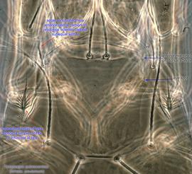
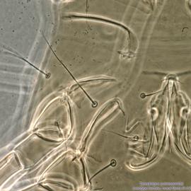
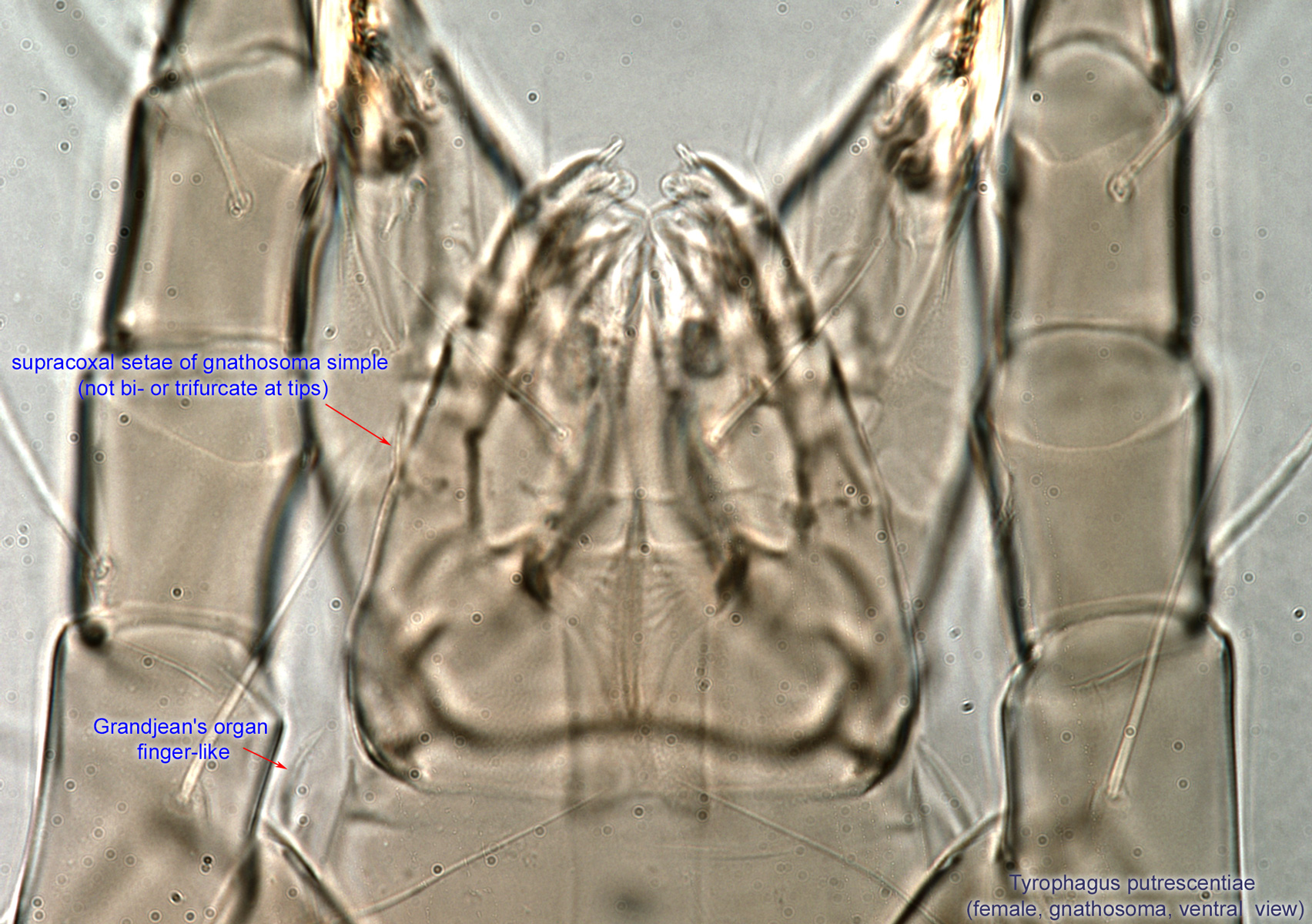
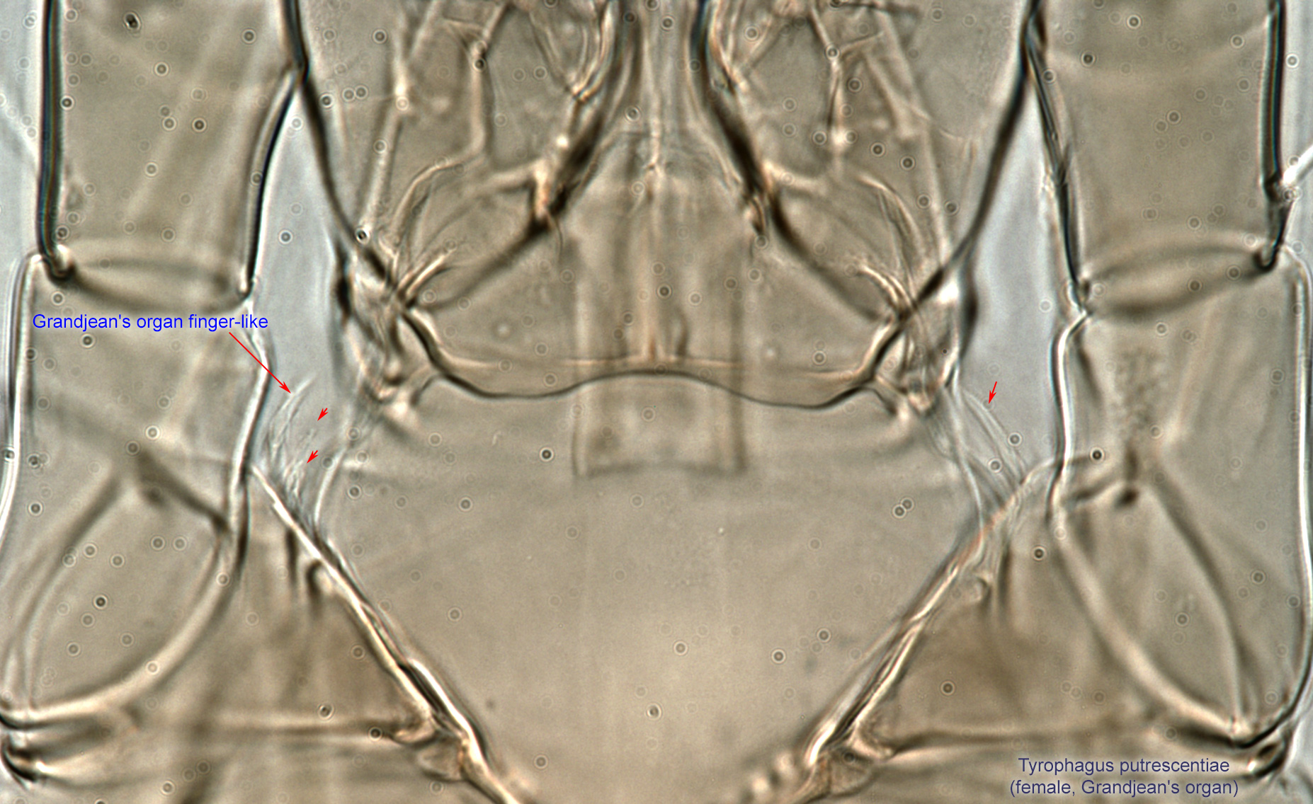
Fig. 6. Tyrophagus putrescentiae female propodosoma, optical section close to ventral side showing Grandjean's organ.
Fig. 6. Tyrophagus putrescentiae female propodosoma, optical section close to ventral side showing Grandjean's organ.
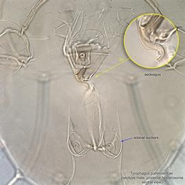
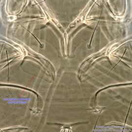
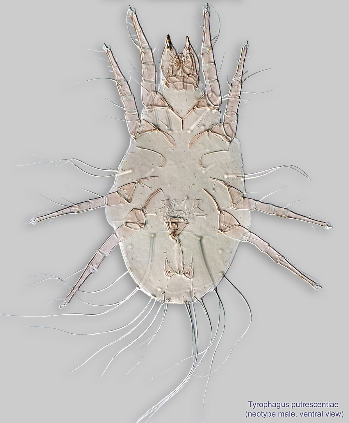
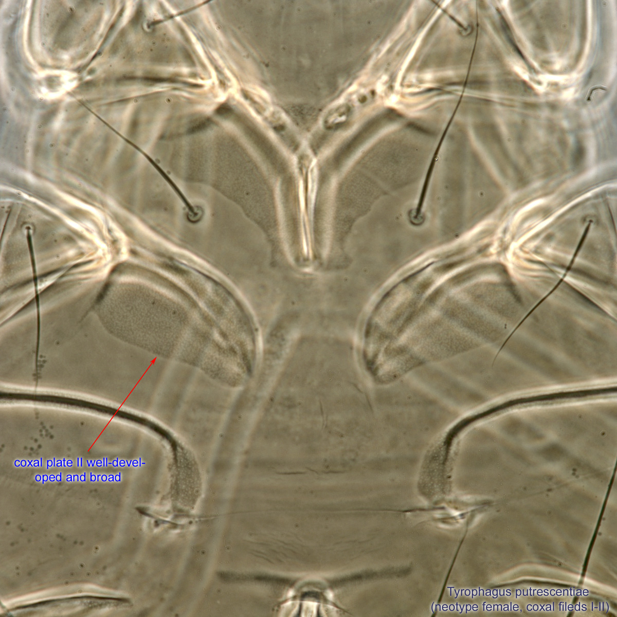
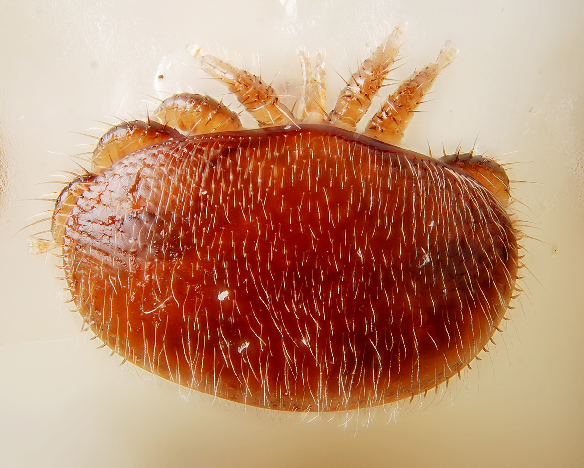
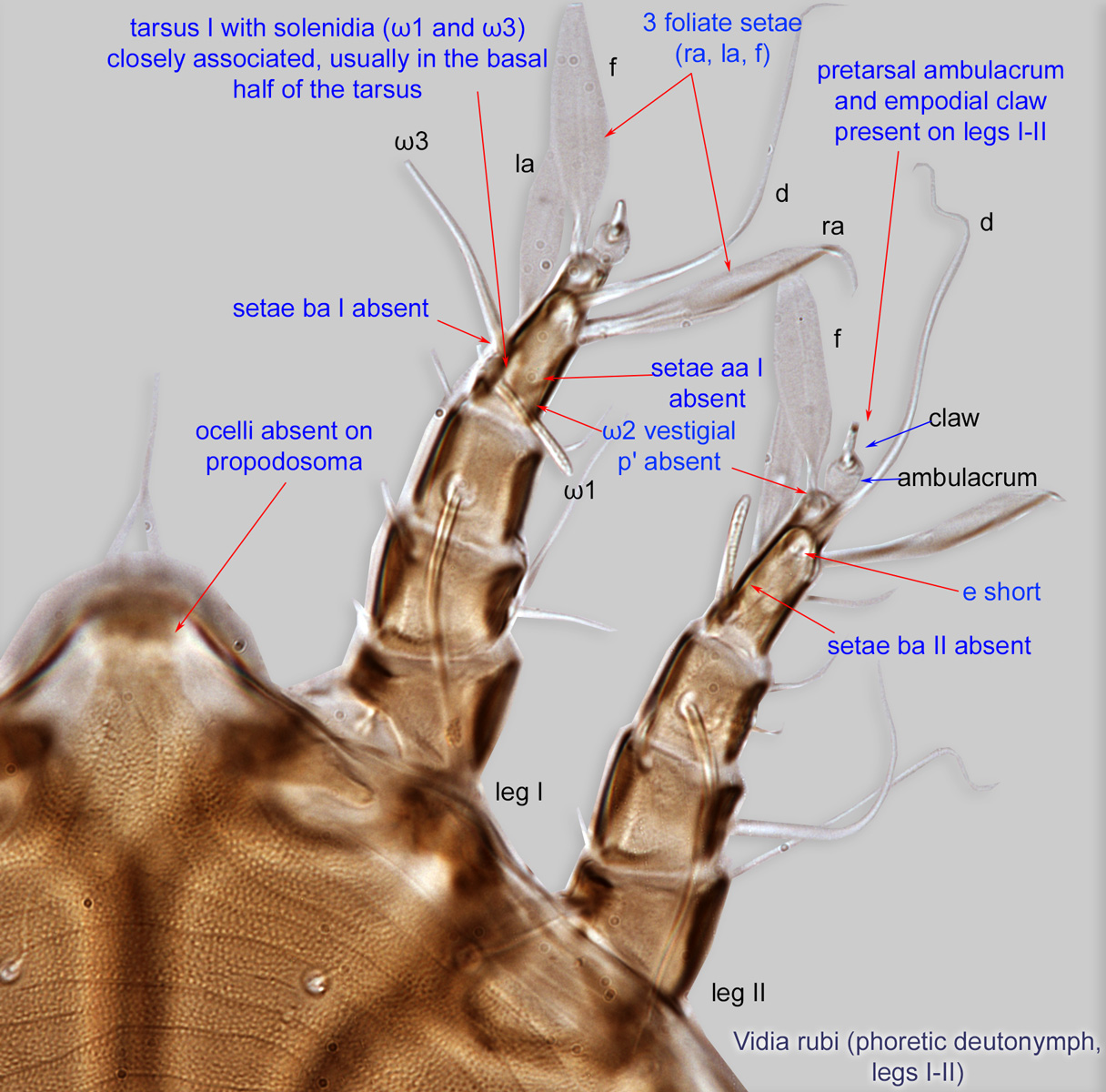
Fig. 13. Tyrophagus putrescentiae female idiosoma showing coxal fields I-II, ventral view.
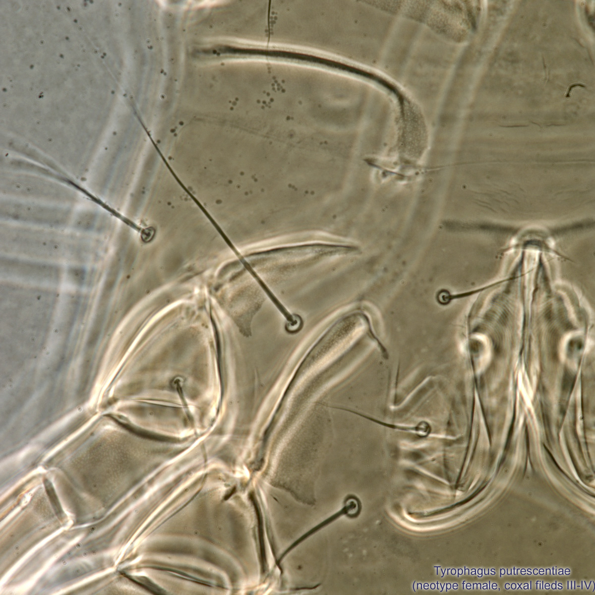
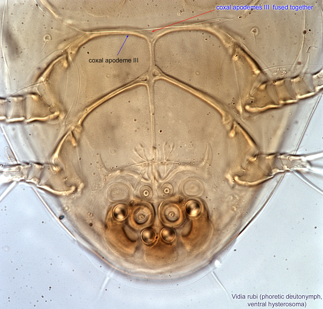
Fig. 14. Tyrophagus putrescentiae female idiosoma showing coxal fields III-IV, ventral view.

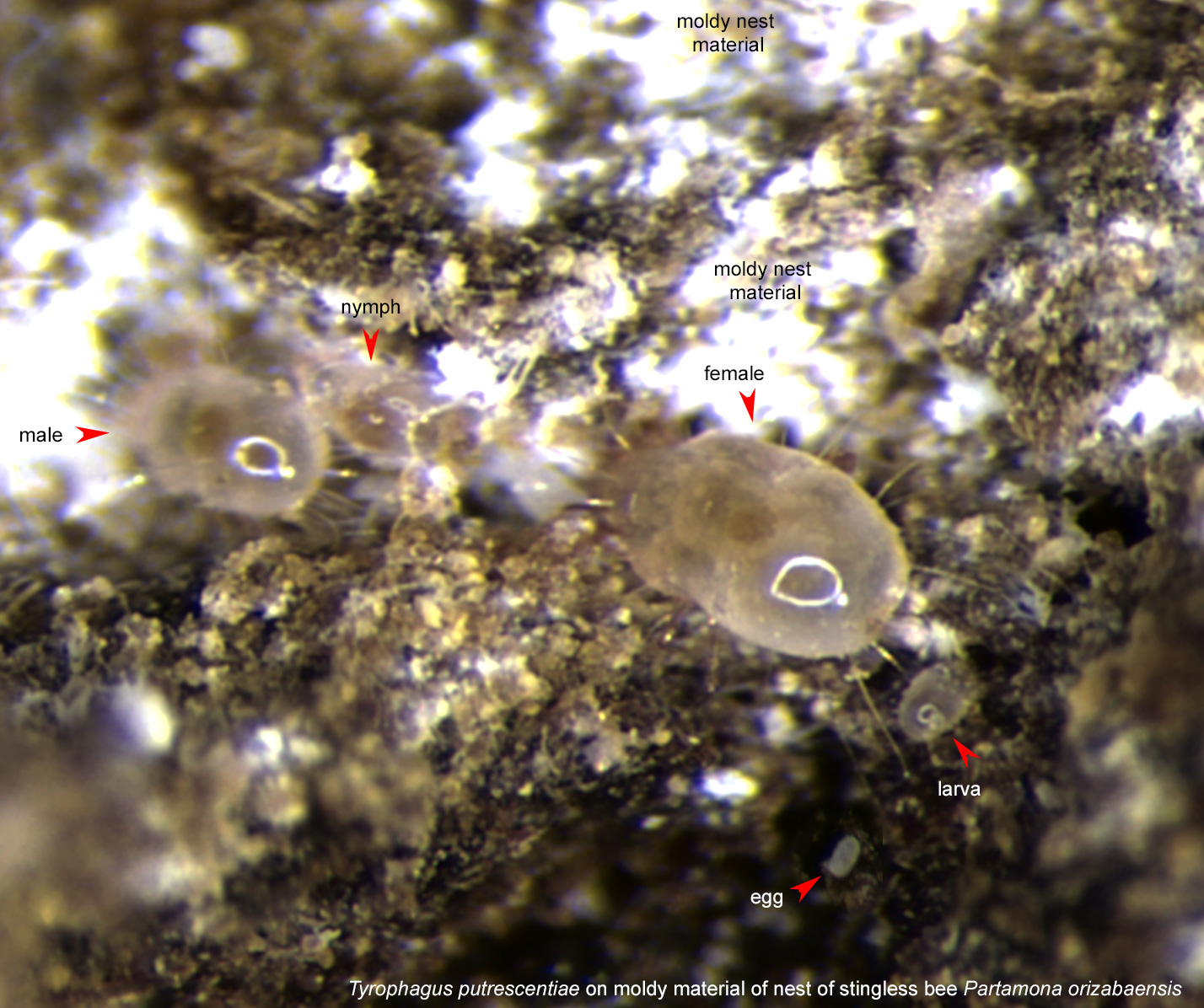
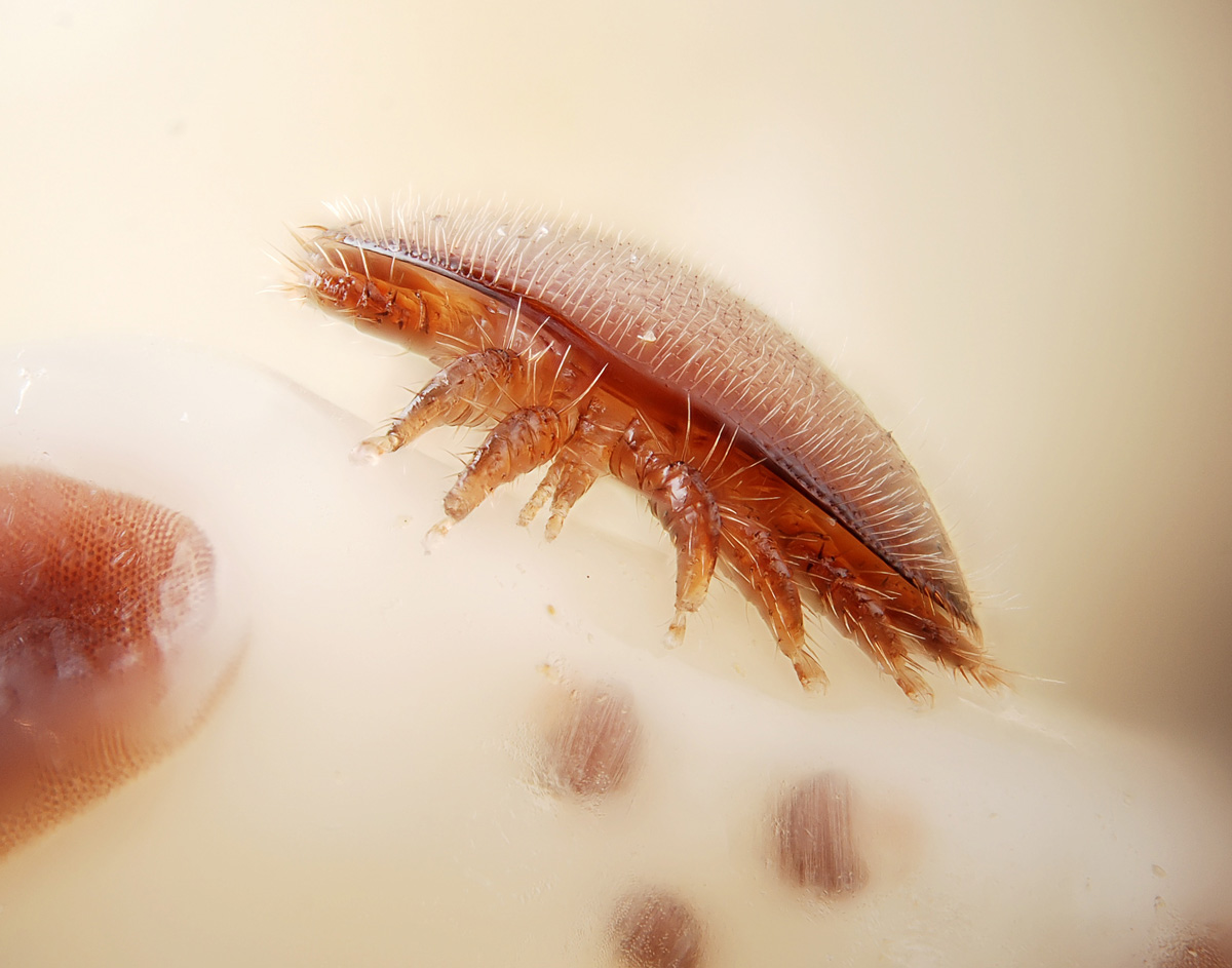
Fig. 15. Tyrophagus putrescentiae on old, moldy material of nest of stingless bee Partamona orizabaensis.
Fig. 15. Tyrophagus putrescentiae on old, moldy material of nest of stingless bee Partamona orizabaensis.
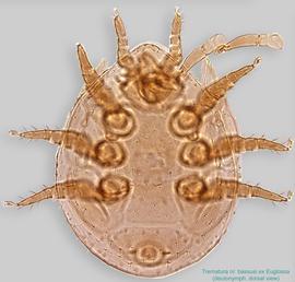
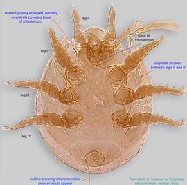
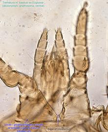
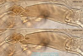
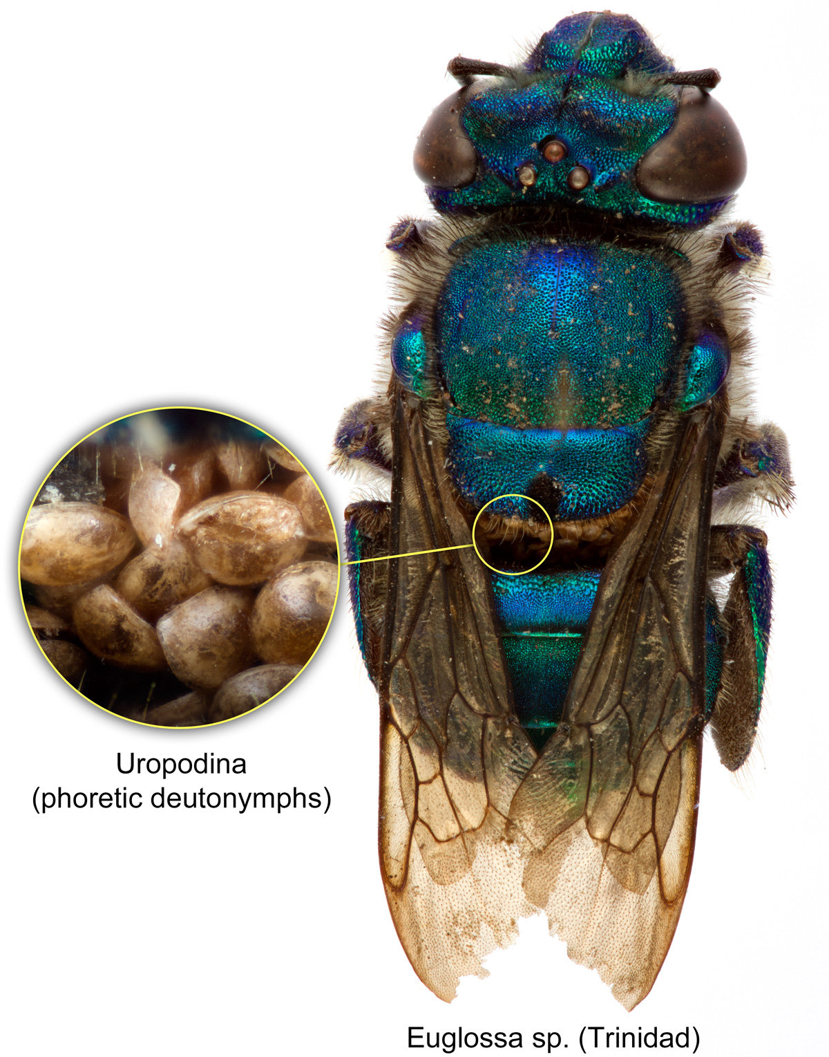
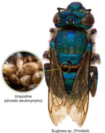 Fig. 5. Uropodina phoretic deutonymphs on bee Euglossa sp. from Trinidad; photo by Lindsey Seastone & Laura Hartmann, ITP.
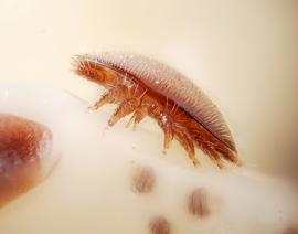
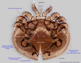
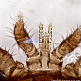
Fig. 4. Female of Varroa destructor parasitizing on the head of pupa of the European honey bee, Apis mellifera; photo by Gilles San Martin/Flickr.
Fig. 4. Female of Varroa destructor parasitizing on the head of pupa of the European honey bee, Apis mellifera; photo by Gilles San Martin/Flickr.
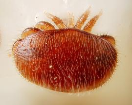
Fig. 5. Female of Varroa destructor on the head of pupa of the European honey bee, Apis mellifera; photo by Gilles San Martin/Flickr.
Fig. 5. Female of Varroa destructor on the head of pupa of the European honey bee, Apis mellifera; photo by Gilles San Martin/Flickr.
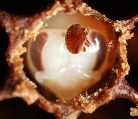
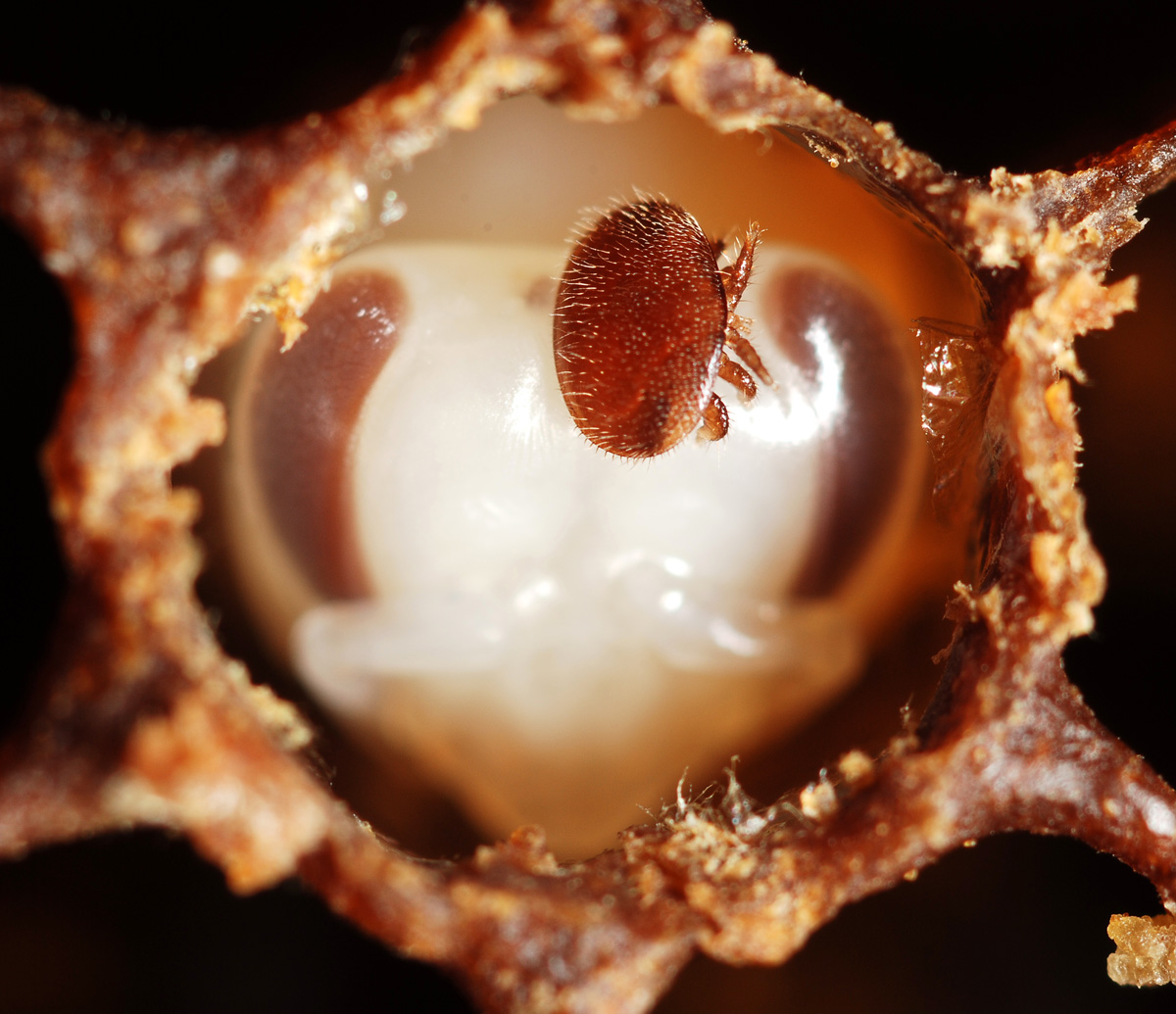
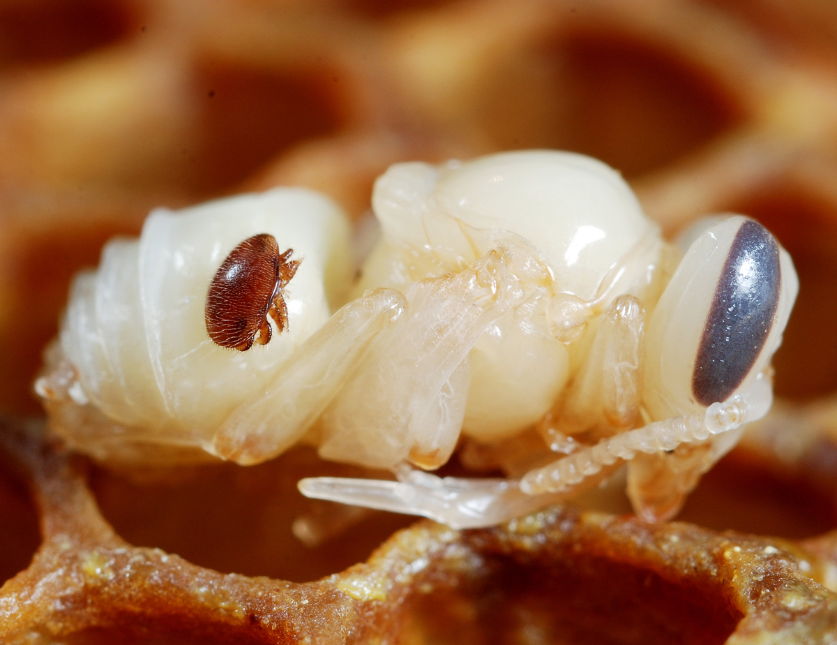
Fig. 6. Female of Varroa destructor on the head of pupa of the European honey bee, Apis mellifera; pupa is inside nest cell; photo by Gilles San Martin/Flickr.
Fig. 6. Female of Varroa destructor on the head of pupa of the European honey bee, Apis mellifera; pupa is inside nest cell; photo by Gilles San Martin/Flickr.
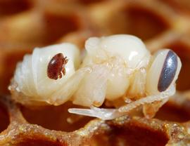
Fig. 7. Female of Varroa destructor on pupa of the European honey bee, Apis mellifera; pupa has been removed from nest cell; photo by Gilles San Martin/Flickr.
Fig. 7. Female of Varroa destructor on pupa of the European honey bee, Apis mellifera; pupa has been removed from nest cell; photo by Gilles San Martin/Flickr.
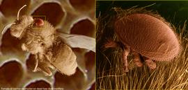
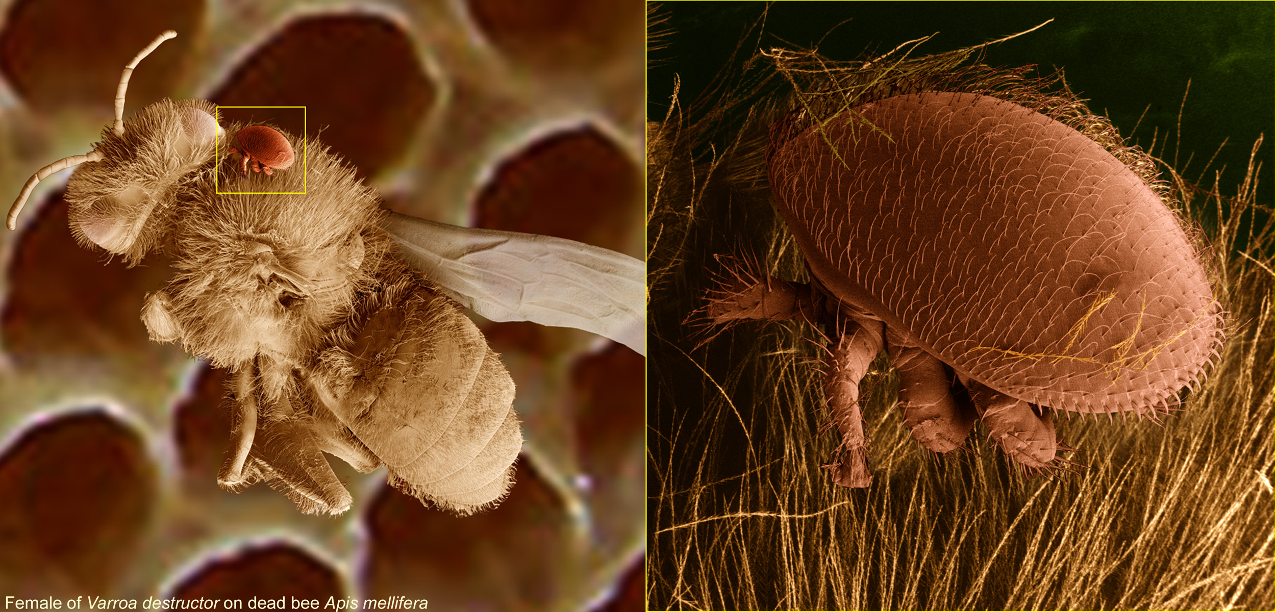
Fig. 8. Female of Varroa destructor on dead adult of the European honey bee, Apis mellifera; LT-SEM (digitally colorized) photo by Ron Ochoa & Gary Bauchan, USDA-ARS.
Fig. 8. Female of Varroa destructor on dead adult of the European honey bee, Apis mellifera; LT-SEM (digitally colorized) photo by Ron Ochoa & Gary Bauchan, USDA-ARS.
Authors: P. Klimov, B. OConnor, R. Ochoa, G. Bauchan, A. Redford, J. Scher
Last updated October 2016
tool images at ITP Node
idtools.org


