

t
mites, ticks
high
Numerous important pests of crops, livestock, wildlife, native flora, and humans.
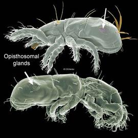
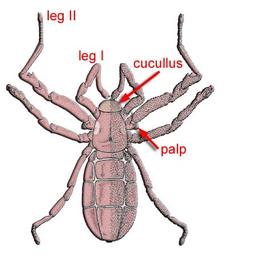
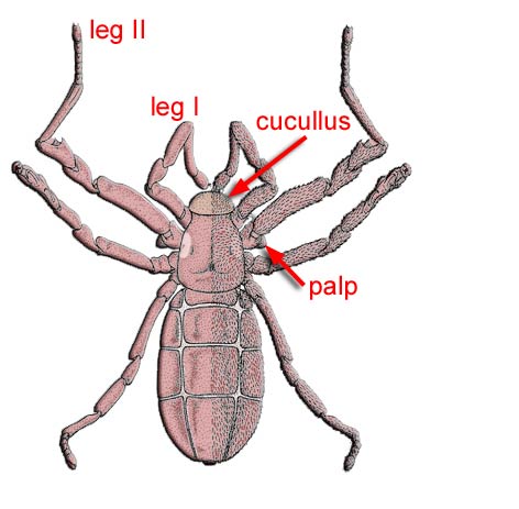
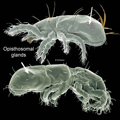
Taxa most similar to mites: Ricinulei have a cucullus and opisthosomal segmentationsegmentation:
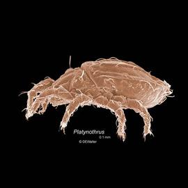
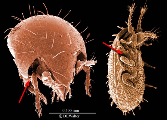
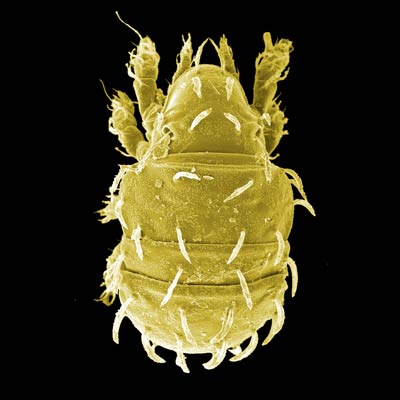
in mites distinct external segments have been lost but remnants of segmentation may be represented by hysterosomal folds or transverse arrays of setae and other cuticular sense organs. In theory, all chelicerates have a prosoma composed of 6 segments (cheliceral, pedipalpal, and four leg-bearing segments = body segments I-VI). Ventrally the positions of the prosomal segments can be identified by the insertions of their appendages, but dorsally they are obscured. The opisthosoma is thought to comprise an additional 12-13 segments (body segments VII-XVIII or XIX), but appears to be somewhat to much reduced in most mites, except possibly Opilioacarida. In early derivative Acariformes (e.g., many Endeostigmata), hysterosomal folds are thought to represent segmentation and in the Grandjean system are designated (from the sejugal furrow to the anus): C, D, E, F, H, PS AD, AN, PA. There is disagreement in the literature over the origin of 'segments' C and D. Adherents of Grandjean consider them to be opisthosomatic (with C probably representing a fusion of the pregenital [body segment VII] and genital [VIII] segments). Others believe that C and D are the dorsal regions of the last two prosomal segments that bear leggs III and IV (i.e., body segments V & VI).
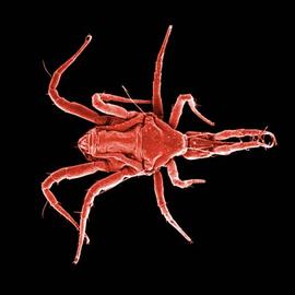
. Opilionids have opisthosomal segmentationsegmentation:
in mites distinct external segments have been lost but remnants of segmentation may be represented by hysterosomal folds or transverse arrays of setae and other cuticular sense organs. In theory, all chelicerates have a prosoma composed of 6 segments (cheliceral, pedipalpal, and four leg-bearing segments = body segments I-VI). Ventrally the positions of the prosomal segments can be identified by the insertions of their appendages, but dorsally they are obscured. The opisthosoma is thought to comprise an additional 12-13 segments (body segments VII-XVIII or XIX), but appears to be somewhat to much reduced in most mites, except possibly Opilioacarida. In early derivative Acariformes (e.g., many Endeostigmata), hysterosomal folds are thought to represent segmentation and in the Grandjean system are designated (from the sejugal furrow to the anus): C, D, E, F, H, PS AD, AN, PA. There is disagreement in the literature over the origin of 'segments' C and D. Adherents of Grandjean consider them to be opisthosomatic (with C probably representing a fusion of the pregenital [body segment VII] and genital [VIII] segments). Others believe that C and D are the dorsal regions of the last two prosomal segments that bear leggs III and IV (i.e., body segments V & VI).
.
See the Mite morphology page for details.
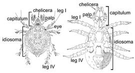
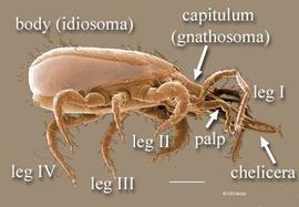
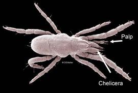
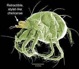
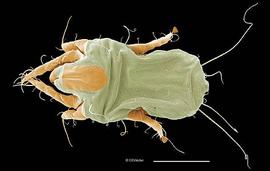
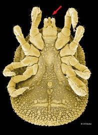
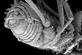
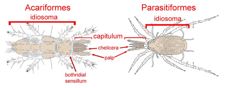
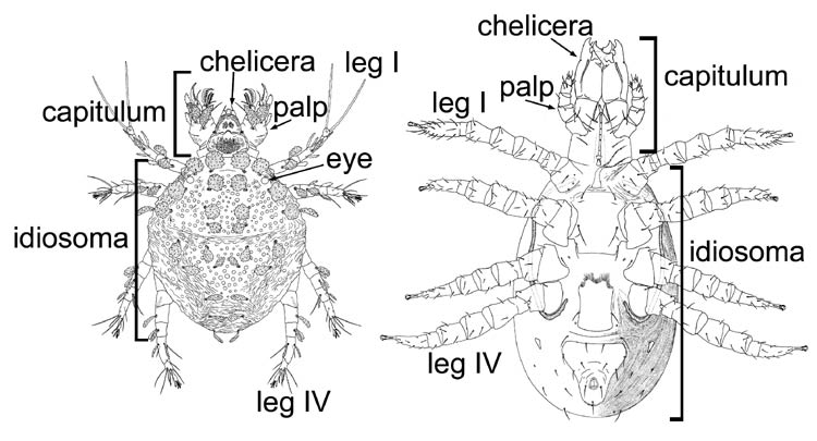
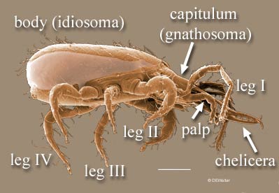
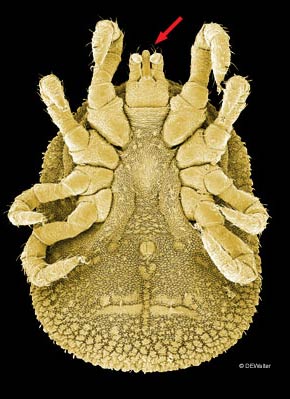
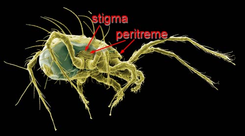
 by flexible cuticle. This anterior region is called the capitulumgnathosoma: is called the idiosoma and lacks primary segmentationsegmentation:
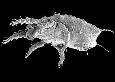 (6 legs); nymphs and adults usually octopod (8 legs).
2–3 superorders, 5–6 orders, 450–500 families: 55,000 described species, >1,000,000 spp.
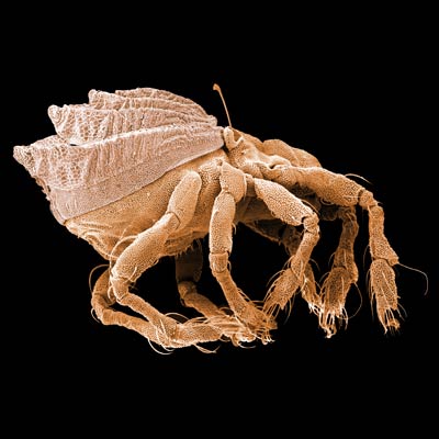
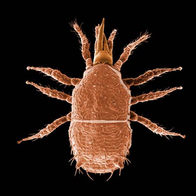
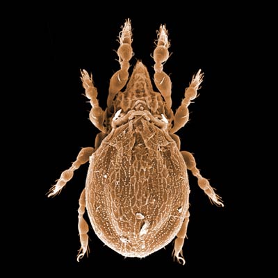
Introduction to the subclass of arachnids that includes mites and ticks
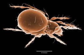
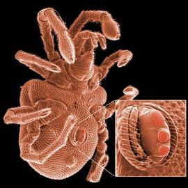
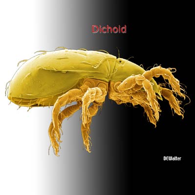
Introduction to the unique morphology of mites
Author: David E. Walter
Content last updated Sept. 2006, unless indicated otherwise
Lucid key player updated Sept. 2025
idtools.org


