Phytophthora parvispora
|
Phytophthora spp. in subclade 7c: portion of the seven-loci ML phylogeny featuring the type cultures of 212 described species (by T. Bourret). Notice the position of P. parvispora Ex-type CBS 132772 = S&T BL 98. Gloria Abad, USDA S&T.
|
|
Phytophthora spp. in subclade 7c: Morphological Tabular key (PDF) and Tabular key legends (PDF) in IDphy2 KEY SECTION. Notice the data of P. parvispora Ex-type CBS 132772 = S&T BL 98. Gloria Abad, USDA S&T.
|
|
Phytophthora parvispora colonies grown for 7 days on (a) V8® Agar, (b) potato dextrose agar, and (c) malt extract agar; photo by Krysta Jennings and Leandra Knight, USDA-APHIS-PPQ |
|
Phytophthora parvispora colony morphology of the ex-type (PH072 – Bruno Scanu) grown for 5 days at 20°C on (a) carrot agar, (b) potato dextrose agar; photos by Bruno Scanu and Antonio Franceschini, Università degli Studi di Sassari, Italy |
|
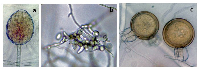
Phytophthora parvispora morphology of ex-type (PH072 – Bruno Scanu) asexual and sexual phases: (a) sporangium, (b) hyphal swellings, (c) gametangia (oogonia and amphigynous antheridia) with plerotic oospores; photos by Bruno Scanu and Antonio Franceschini, Università degli Studi di Sassari, Italy |
|
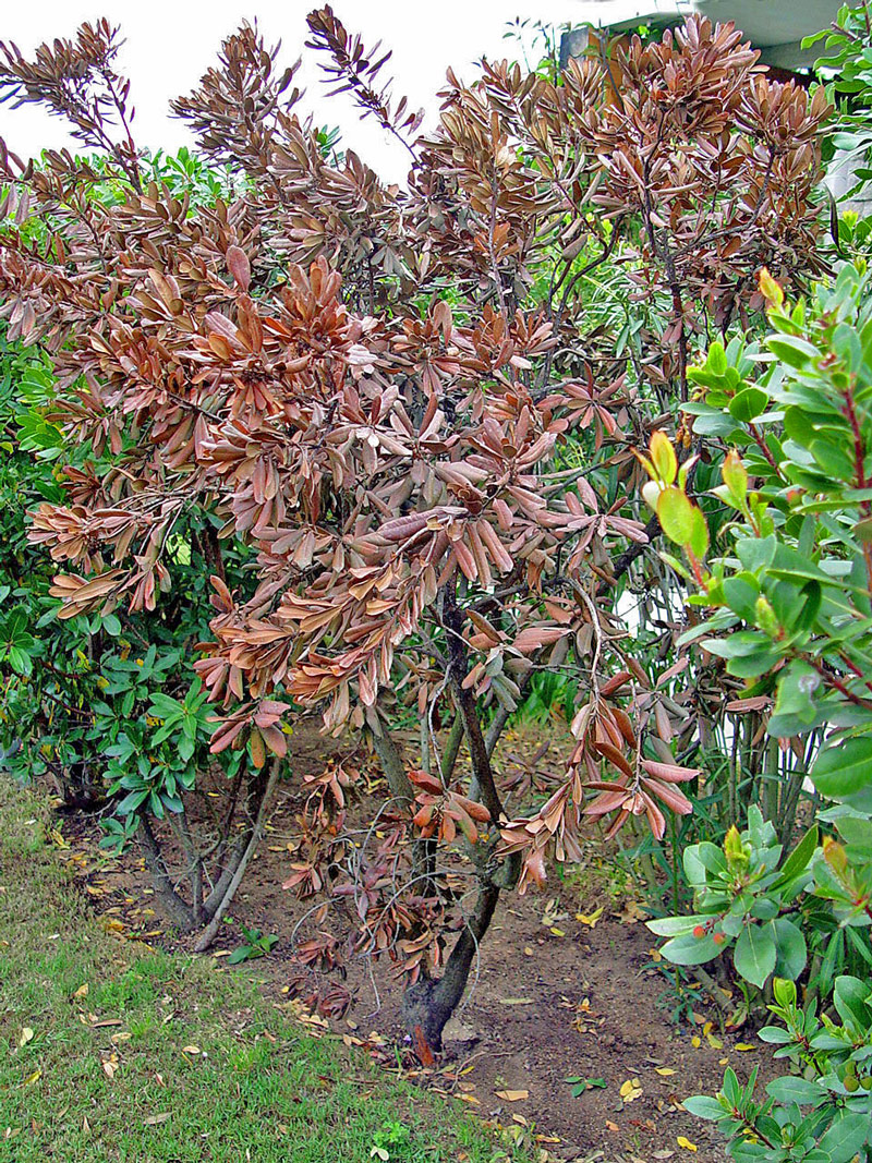
Arbutus unedo in an ornamental planting showing symptoms caused by Phytophthora parvispora in Sardinia, Italy; photo by Bruno Scanu and Antonio Franceschini, Università degli Studi di Sassari, Italy |
|
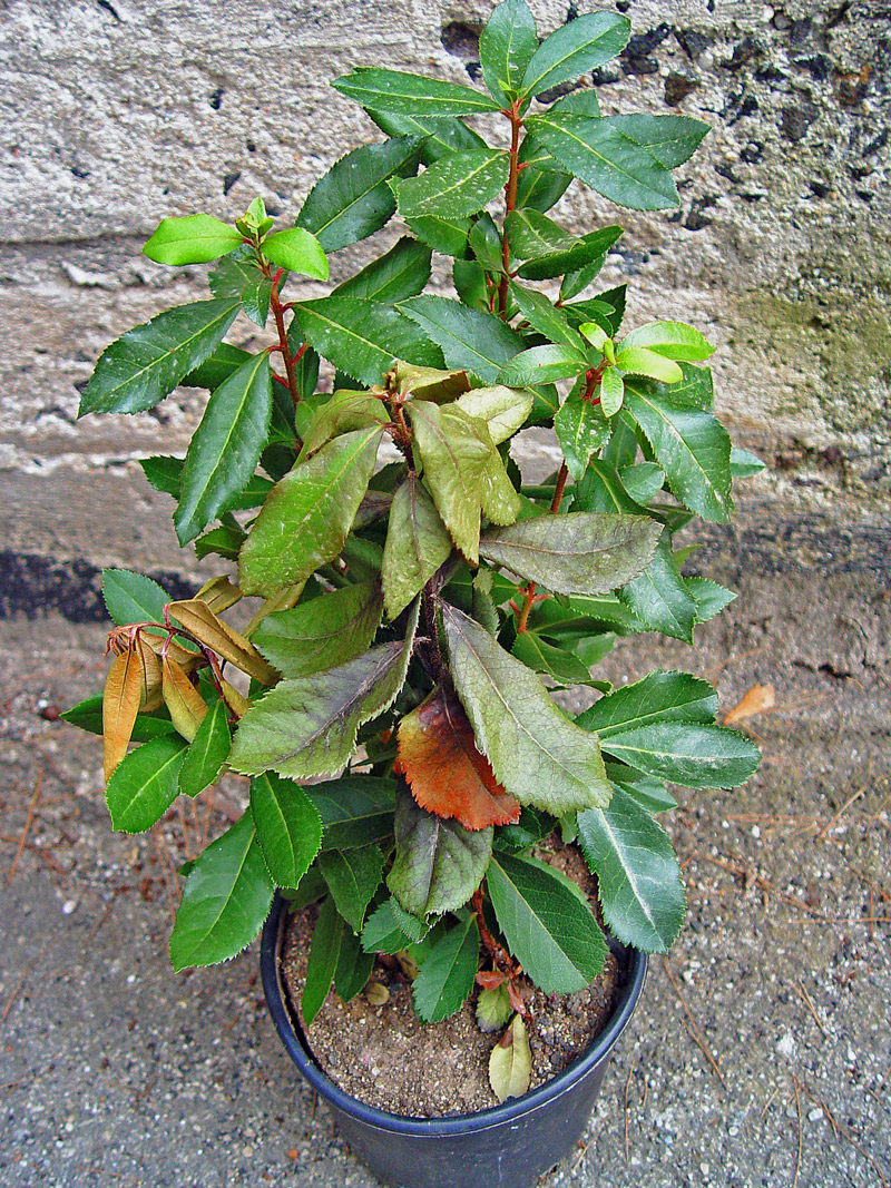
Arbutus unedo in nursery with shoot dieback and wilting caused by Phytophthora parvispora in Sardinia, Italy; photo by Bruno Scanu and Antonio Franceschini, Università degli Studi di Sassari, Italy |
Name and publication
Phytophthora parvispora Scanu and Denman (2014)
Scanu B, Hunter GC, Linaldeddu BT, Franceschini A, Maddau L, Jung T, and Denman S. 2014. A taxonomic re-evaluation reveals that Phytophthora cinnamomi and Phytophthora cinnamomi var. parvispora are separate species. Forest Pathol. 44: 1–20.
Corresponding author: bscanu@uniss.it
Nomenclature
from Scanu et al. (2014)
Mycobank
Synonym
= Phytophthora cinnamomi var. parvispora Kröber & Marwitz, Zeitschrift für Pflanzenkrankheiten und Pflanzenschutz 100: 255 (1993) [MB360187]
Etymology
refers to the small-sized reproductive structures
Typification
Type: ITALY, Sardinia, isolated from collar rot of a declining planted Arbutus unedo collected by B. Scanu, 2011; CBS H-21132 (holotype, dried culture on CA, Herbarium CBS-KNAW Fungal Biodiversity Centre)
Ex-type: CBS 132772 = PH072 (culture ex-type A1)
Sequences for ex-type in original manuscript: CBS 132772 = ITS KC478667, β-tub KC609402, Cox1 KC609413 (FM 85/84), Cox2 KC559763 (FM 75/78)
Ex-type in other collections
(ET) CBS 132772, NRRL 64106, WPC P19847 (A2), S&T BL 98 (Abad), PH072 (Scanu), TJ 1542 (Jung), 66C8 (Hong)
Molecular identification
Voucher sequences for barcoding genes (ITS rDNA and COI) of the ex-type (see Molecular protocols page)
Phytophthora parvispora isolate CBS 132772 = ITS rDNA KC478667
Phytophthora parvispora isolate CPHST BL 98 ( = P19847) COI MH136953
Sequences for selected specimens
Phytophthora parvispora isolate CBS 411.96 = ITS rDNA KC478672
Phytophthora parvispora isolate CPHST BL 107 ( = P8495 ) COI MH136955
Voucher sequences for Molecular Toolbox with seven genes (ITS, β-tub, COI, EF1α, HSP90, L10, and YPT1
(see Molecular protocols page) (In Progress)
Voucher sequences for Metabarcoding High-throughput Sequencing (HTS) Technologies [Molecular Operational Taxonomic Unit (MOTU)]
(see Molecular protocols page) (In Progress)
Sequences with multiple genes for ex-type in other sources
- NCBI: Phytophthora parvispora CPHST BL 98
- NCBI: Phytophthora parvispora CBS 132772
- EPPO-Q-bank: Phytophthora parvispora CBS 132772
- BOLDSYSTEMS: Phytophthora parvispora (barcoding COI & ITS)
Position in multigenic phylogeny with 7 genes (ITS, β-tub, COI, EF1α, HSP90, L10, and YPT1)
Clade clade:
a taxonomic group of organisms classified together on the basis of homologous features traced to a common ancestor
7c
Morphological identification
adapted from Scanu et al. (2014)
Colonies and cardinal temperatures
Colonies on V-8 agar slight chrysanthemum to stellate patten, in potato dextrose agar and malt extract agar with slow growth, in carrot agar with chrysanthemum to stellate pattern. Minimum temperature for growth 10°C, optimum 27°C, maximum 37°C.
Conditions for growth and sporulation
Sporangia are abundantly produced in liquid cultures after 24 hrs.
Asexual phase
SporangiaSporangia:
sac within which zoospores form, especially when water is cooled to about 10°C below ambient temperature; in solid substrates, sporangia usually germinate by germ tubes
nonpapillatenonpapillate:
pertaining to the production of a non-distinct, or inconspicuous, papilla at the distal end of the sporangium (cf. papillate and semipapillate)
; persistentpersistent:
pertaining to sporangia that remain attached to the sporangiophore and do not separate or detach easily (cf. caducous)
; ovoidovoid:
egg-shaped, with the widest part at the base of the sporangium and the narrow part at the apex
, ellipsoidellipsoid:
refers to a solid body that forms an ellipse in the longitudinal plane and a circle in cross section; many fungal spores are ellipsoidal or elliptic
or obpyriformobpyriform:
inversely pear-shaped, i.e. with the widest part at the point of attachment (cf. pyriform)
(31–43 µm L x 20–26 µm W); showing nested and extended internal proliferationinternal proliferation:
internal proliferation occurs when the sporangiophore continues to grow through an empty sporangium
, frequently producing long chains of proliferating sporangiasporangia:
sac within which zoospores form, especially when water is cooled to about 10°C below ambient temperature; in solid substrates, sporangia usually germinate by germ tubes
along the same sporangiophoresporangiophore:
the hyphal strand on which the sporangium is formed; may be branched or unbranched to form compound sympodia or simple sympodia
, external proliferationexternal proliferation:
formation of a sporangium after a sporangiophore has emerged from beneath and external to an empty sporangium that has previously emitted its zoospores (cf. internal proliferation)
is also observed; sporangiasporangia:
sac within which zoospores form, especially when water is cooled to about 10°C below ambient temperature; in solid substrates, sporangia usually germinate by germ tubes
originated mostly in unbranched sporangiophores and sometimes intercalaryintercalary:
positioned within a hypha (cf. terminal)
. Hyphal swellings irregular or globoseglobose:
having a rounded form resembling that of a sphere
, often formed in cluster aggregations. ChlamydosporesChlamydospores:
an asexual spore with a thickened inner wall that is delimited from the mycelium by a septum; may be terminal or intercalary, and survives for long periods in soil
occasionally formed and are globoseglobose:
having a rounded form resembling that of a sphere
(19-24 µm diam.) and produced mostly laterally and rarely terminally.
Sexual phase
Heterothallicheterothallic:
pertaining to sexual reproduction in which conjugation is possible only through interaction of different thalli (i.e. different mating types) (cf. homothallic)
. OogoniaOogonia:
the female gametangium in which the oospore forms after fertilization by the antheridium
golden brown, smooth-walled and globoseglobose:
having a rounded form resembling that of a sphere
(31–33 µm diam.); antheridiaantheridia:
the male gametangium; a multinucleate, swollen hyphal tip affixed firmly to the wall of the female gametangium (the oogonium)
amphigynousamphigynous:
pertaining to the sexual stage in which the antheridium completely surrounds the stalk of the oogonium (cf. paragynous)
and globoseglobose:
having a rounded form resembling that of a sphere
; oosporesoospores:
zygote or thick-walled spore that forms within the oogonium after fertilization by the antheridium; may be long-lived
apleroticaplerotic:
pertaining to a mature oospore that does not fill the oogonium; i.e. there is room left between the oospore wall and oogonium wall (cf. plerotic)
to slightly apleroticaplerotic:
pertaining to a mature oospore that does not fill the oogonium; i.e. there is room left between the oospore wall and oogonium wall (cf. plerotic)
(25–26 µm diam.).
Most typical characters
Phytophthora parvispora is very similar to P. cinnamomi in the morphological characters of the asexual phase. Phytophthora parvispora produces occasionally small chlamydosporeschlamydospores:
an asexual spore with a thickened inner wall that is delimited from the mycelium by a septum; may be terminal or intercalary, and survives for long periods in soil
in comparison to the frequent chlamydosporeschlamydospores:
an asexual spore with a thickened inner wall that is delimited from the mycelium by a septum; may be terminal or intercalary, and survives for long periods in soil
produced by P. cinnamomi.
Additonal specimen(s) evaluated
Phytophthora parvispora ex-type CPHST BL 98 (A1), duplicate of P19847 (World Oomycetes/Phytophthora Collection), which is a duplicate of CBS 132772
CPHST BL 13 = P8494 WPC A1 from Beaucamea sp., collected by R. Marwitz in Germany, Frankfurt in 1990 = CBS 411.96 (BBA 65507)
CPHST BL 107= P8495 WPC A2 from Beaucamea sp., collected by R. Marwitz in Germany, Frankfurt in 1990 = CBS 413.96 (BBA 65508)
Hosts and distribution
Distribution: Europe (Germany, Italy, Portugal), South Africa, South America (Brazil), Taiwan, possibly also Australia
Substrate: roots and stems
Disease note: collar and stem rot, dieback
Host: diverse hosts
Retrieved February 01, 2018 from U.S. National Fungus Collections Nomenclature Database.
Additional Info
Hosts: Arbutus unedo, Mandevilla sp., Pinus pinea, Beaucamea sp., and Agathosma betulina
Quarantine status
USA Status: This species was listed as a species of concern during the 2009 Phytophthora prioritization project conducted by USDA APHIS PPQ CPHST PERAL (Schwartzburg et al.).
Phytophthora parvispora is also listed in the U.S. Regulated Plant Pest Table (last modified Nov. 15, 2017).
Additional references and links
- SMML USDA-ARS: Phytophthora parvispora
- EPPO Global Database: Phytophthora parvispora
- Forest Phytophthoras of the World: Phytophthora parvispora
- CABI Digital Library: Phytophthora parvispora
- Encyclopedia of Life (EOL): Phytophthora parvispora
- Index Fungorum (IF): Phytophthora parvispora
- Google All Phytophthora parvispora
- Google Images Phytophthora parvispora
- Google Scholar Phytophthora parvispora
see also Phytophthora cinnamomi parvispora
Fact sheet author
Z. Gloria Abad, Ph.D., USDA-APHIS-PPQ-S&T Plant Pathogen Confirmatory Diagnostics Laboratory (PPCDL), United States of America.


