
Adoxophyes negundana (McDunnough) (Tortricidae: Tortricinae: Archipini)
FWL: 7.5–9.5 mm
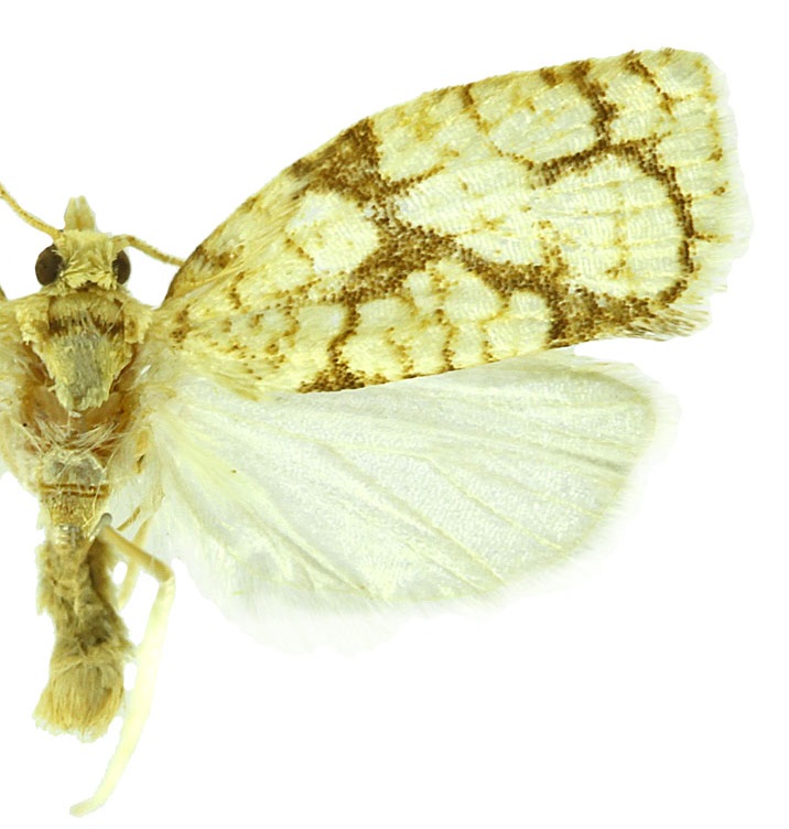
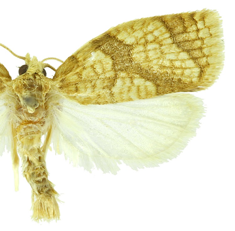
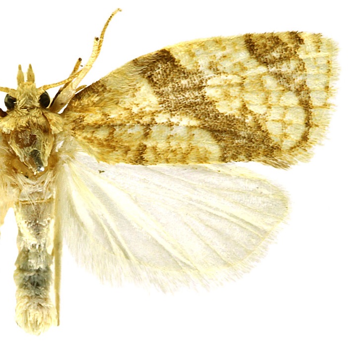
Adults are pale yellow with light brown fasciae and a network of light brown streaks along and between veins. Hindwings are white. Males have a forewing costal fold.
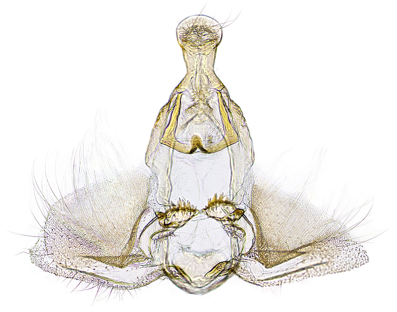
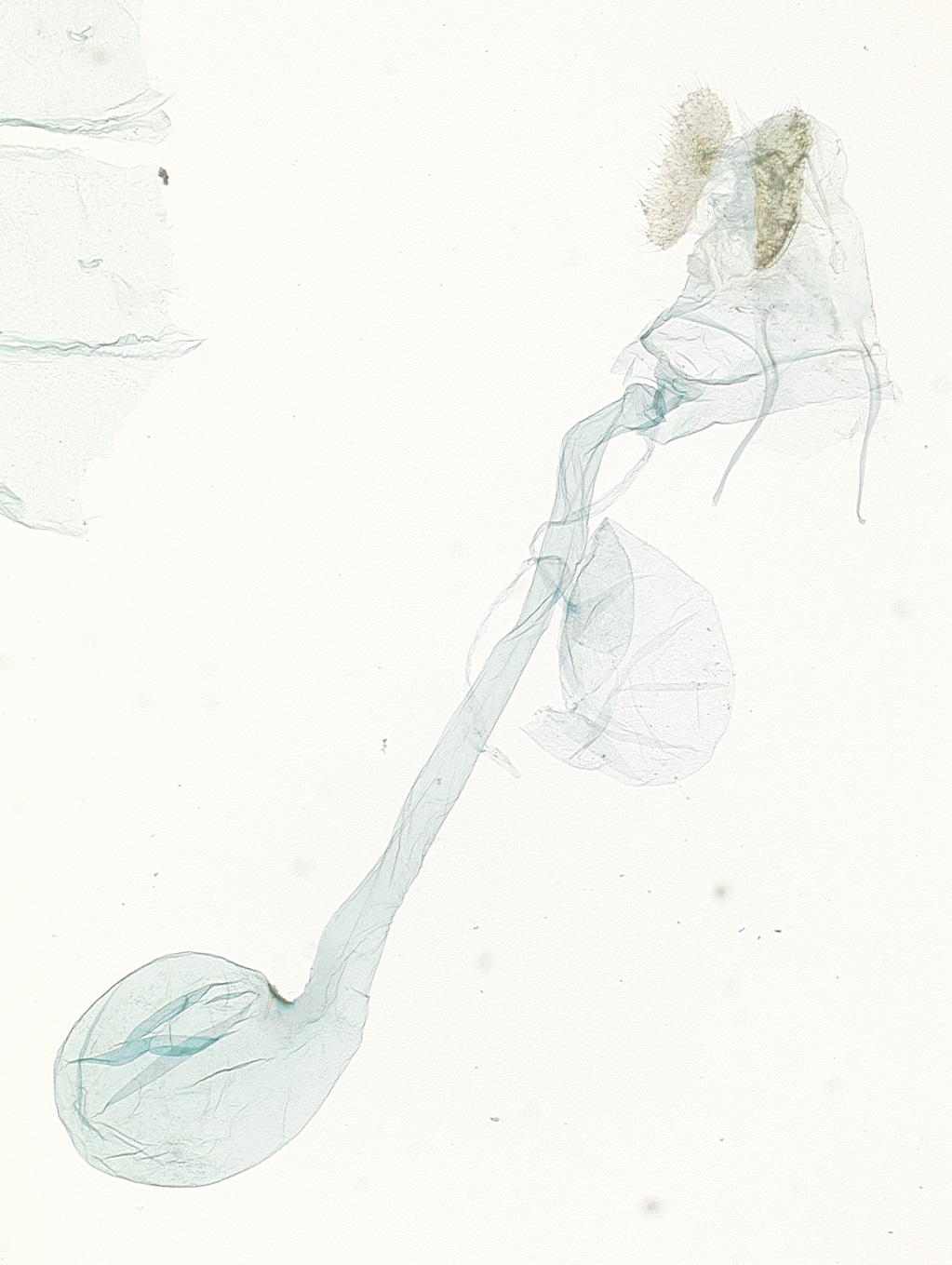
Male genitalia are characterized by a spatulate uncus; an incomplete, finely spined transtilla; and large, rounded membranous valvae. Female genitalia have not been described in the published literature, but closely resemble those of Adoxophyes furcatana.
Larval morphology is undocumented for this species.
Adoxophyes negundana and A. furcatana are the only two representatives of the genus Adoxophyes in North America. These two species are difficult to separate using forewing pattern. In general, the median fascia is broader in A. negundana, although this character does not appear to be consistent across all individuals.
Freeman (1958)Freeman (1958):
Freeman, T. N. 1958. The Archipinae of North America (Lepidoptera: Tortricidae). Canadian Entomologist, Supplement 7 (Vol. 90): 1-89. states that male genitalia of A. negundana have fewer cornuti (only four) and more sharply elbowed gnathos arms than those of A. furcatana. Both species of Nearctic Adoxophyes are similar to A. orana and might be confused with this Palearctic species if it were discovered in North America. It is not known if Nearctic Adoxophyes are attracted to A. orana pheromone.
Adoxophyes negundana is found from New Brunswick to Manitoba, south to Florida and west to Utah.
The life history of this species is poorly known. Adults are present from June through early September. Larvae feed in the rolled leaves of boxelder (Acer negundo).
View full screen host table here
Authors: KA Austin, TM Gilligan, ME Epstein
Content last updated May 2025
idtools.org
TortAI tool images at ipmimages.org - ITP Node